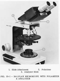
Polarized light is made available for the
qualitative examination of crystals, fibers,
minerals, etc. with the use of the Polarizer,
Cat. No. 31-57-77. This Polarizer fits in
the filter recess of the High Intensity Base
Illuminator and the Professional Optilume
and fits over the lens of the Optilume,
Fig. 13-1.
Two types of Analyzers are available. An Eyecap Analyzer, Cat. No. 31-57-84, may be used over the Eyepiece, or an Analyzer Slide, Cat. No. 31-57-86, may be used in conjunction with the Slide Attachment, Cat. No. 31-57-92, Fig. 13-2.
The Eyecap Analyzer and Polarizer combine to form an economical means of providing Polarized Light Microscopy. They are best suited to elementary work and highly birefringent specimens, which give a high contrast in polarized light.
In the fabrication of the optics a slight
amount of residual strain may result. This
amount of strain will not affect the image
quality in other types of microscopy. The
effect in Polarized Light Microscopy may
be a lowering of the contrast in comparison
to that of Polarizing Microscopes using
strain-free optics. The amount of residual
strain may be lessened by using fewer optics
between the Polarizer and Analyzer. For
this reason it is recommended that the
Analyzer Slide be used thus eliminating
the effect of the optics in the Microscope
Head. If the specimen is highly birefringent,
the effect of this strain will be
of no consequence.
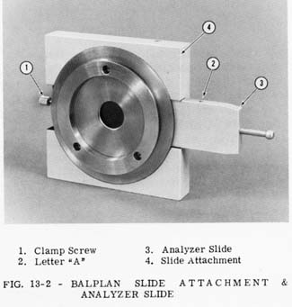
The Analyzer Slide and Slide Attachment may be installed on your Balplan Microscope as follows:
When in this orientation, the vibration axis of the Analyzer will be parallel to the long dimension of the Microscope Arm.
The Analyzer Slide has the letter "A" engraved on one side of the slide. Fig. 13-2. In the opposite side of the slide are two slots that are part of the detent mechanism. The slide must be inserted in the slot of the Slide Attachment, with the letter "A" facing you, in order for the detents to function. When the Analyzer is in the correct detent position for Polarized Light Microscopy, the letter "A" will be visible. In the other detent position, the letter "A" is not visible.
The Eyecap Analyzer has an index line which represents the axis of vibration. When it is installed over the Eyepiece, it should be oriented either perpendicular or parallel to an imaginary line connecting the centers of the Eyepieces.
The Polarizer must be oriented so that its transmission axis is perpendicular to the axis of the Analyzer. The index line on the Polarizer is parallel to the Polarizer's axis.
With the Analyzer installed as directed, place the Polarizer in the filter recess or over the lens of the Illuminator. While observing through the Eyepiece, rotate the Polarizer until extinction (minimum brightness) occurs.